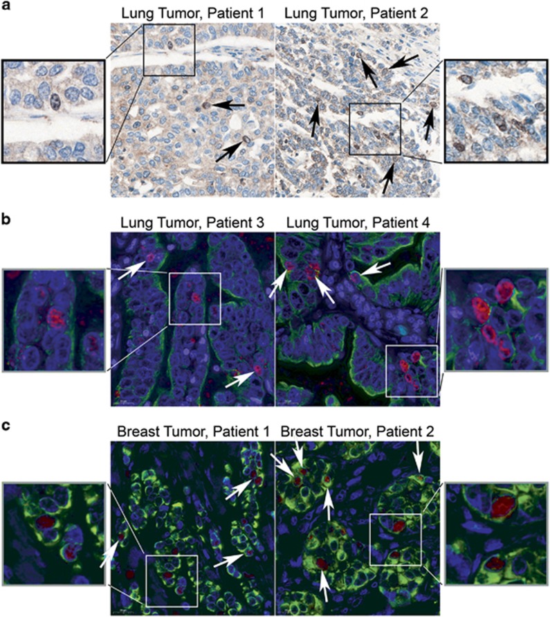
Figure 7.
Nuclear SmgGDS is detectable in patients’ lung and breast tumors. (a) Representative immunohistochemical staining of SmgGDS in patients’ lung tumors is shown. Black arrows indicate cells with nuclear and nucleolar SmgGDS. (b, c) Representative immunofluorescent staining of SmgGDS (red) and cytokeratin (green) in patients’ lung tumors (b) and breast tumors (c) is shown. White arrows indicate cells with nuclear and nucleolar SmgGDS.

