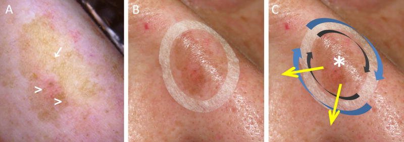
Figure 1.
Lentigo maligna margin determination using dermoscopy, Wood´s lamp examination and handheld reflectance confocal microscopy. A, Dermoscopy evaluation showing a pigmented lesion with ill-defined margins, asymmetric pigmented follicular openings (arrow) and circle within a circle (arrowheads). B, Paper ring placed outside the clinical margin determined with dermoscopy and Wood’s lamp to facilitate confocal navigation. C, Handheld reflectance confocal microscopy evaluation was performed by imaging initially the center to determine the cell morphology (asterisk), and later by imaging clockwise the peripheral margin inside the ring (grey arrows) and outside the ring (blue arrows). Radial videos were obtained (yellow arrows) in the areas where a higher degree of atypia was identified outside the paper ring to determine the LM/LMM subclinical extension.

