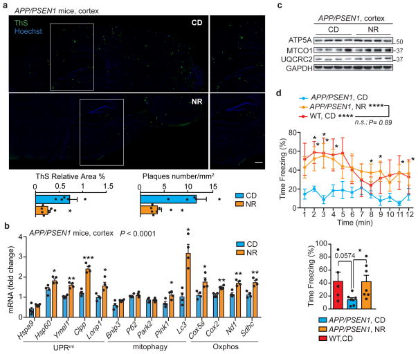
Figure 5. NR reduces Aβ deposits, induces the MSR and improves contextual memory in transgenic AD mice.
a, Representative images and corresponding quantification of plaques in cortex samples of APP/PSEN1 AD mice following NR treatment, stained using Thioflavin S (ThS) (CD, n=5 animals; NR, n=7 animals; NR 400mg/kg/day for 10 weeks). Scale bar, 200μm. *P<0.05 (relative % area,0.041; number,0.014). b–c, MSR transcript (b) and immunoblot (c) analyses of cortex samples of APP/PSEN1 mice following NR treatment (b, n=5 animals per group; c, n=4 animals per group). Data in b,c are representative of two independent experiments. d, Contextual fear conditioning in WT (n=5 animals) and APP/PSEN1 mice with or without NR treatment (n=7 animals), plotted in function of the time intervals (left) or as average of the total obtained values (right). *P<0.05 (P=0.03); ****P≤0.0001. One-tail t test performed between WT and APP/PSEN1 averaged values (P=0.0574, 95% confidence interval). Values in the figure are mean ± s.e.m. *P<0.05; **P≤0.01; ***P≤0.001; ****P≤0.0001; n.s., non-significant. Throughout the figure, overall differences between conditions were assessed by two-way ANOVA. Differences for individual genes or two groups were assessed using two-tailed t tests (95% confidence interval). See Methods for further details. CD, chow diet. For uncropped gel source data, see Supplementary Fig. 1. For all the individual p values, see the Fig. 5 Spreadsheet file.

