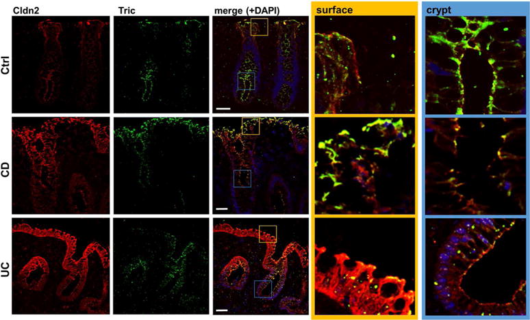
Fig. 2. Localization of claudin 2 (red) and tricellulin (green) in human colon biopsies.

Representative immunofluorescent stainings of cryosections of patient biopsies. While claudin-2 is only expressed within the crypts of control patients (ctrl), it is expressed all along the crypt and surface in CD and UC patients. Tricellulin is present in all areas of the crypt and is also detectable in surface areas of the control patients. In UC, a decreased expression is observed, while in CD patients there seems to be a shift of expression; while in the crypts the signal appears to be reduced, localization within the surface is increased. The merged localizations within the crypt (blue box) and the surface epithelium (yellow) are also shown in higher magnifications. Bar = 50 nm.
