Figure 1.
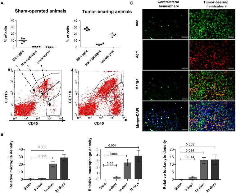
Evaluation of the content of microglia, blood-derived macrophages and leukocytes in C6 rat gliomas. (A) Percentages of microglia, blood-derived macrophages and leukocytes in brains of sham-operated and C6 rat glioma bearing animals at day 21st after implantation. Lower panel shows gating strategy and representative results of FACS analysis. Quadrant gates were drawn on three cell subpopulations based on differences in the surface expression of CD11b and CD45 antigens: microglia (CD11b+CD45low), blood-derived macrophages (CD11b+CD45high) and leukocytes (CD11b-CD45high). Leukocytes CD11b-CD45high and CD11b+CD45high invading macrophages/monocytes were mostly absent in sham-operated brain samples. (B) Kinetics of accumulation of microglia, peripheral macrophages and leukocytes within the tumor. Changes in cell type content at 8th, 14th and 21st day after implantation were calculated as content of each subpopulations in glioma bearing brains related to its content in sham-operated animals (N = 4–6 per time point). (C) Double staining of Iba1 + and Arginase 1 shows accumulation and the pro-tumorigenic activation of GAMs only in tumor-bearing hemispheres (N = 3–5 rats per group).

