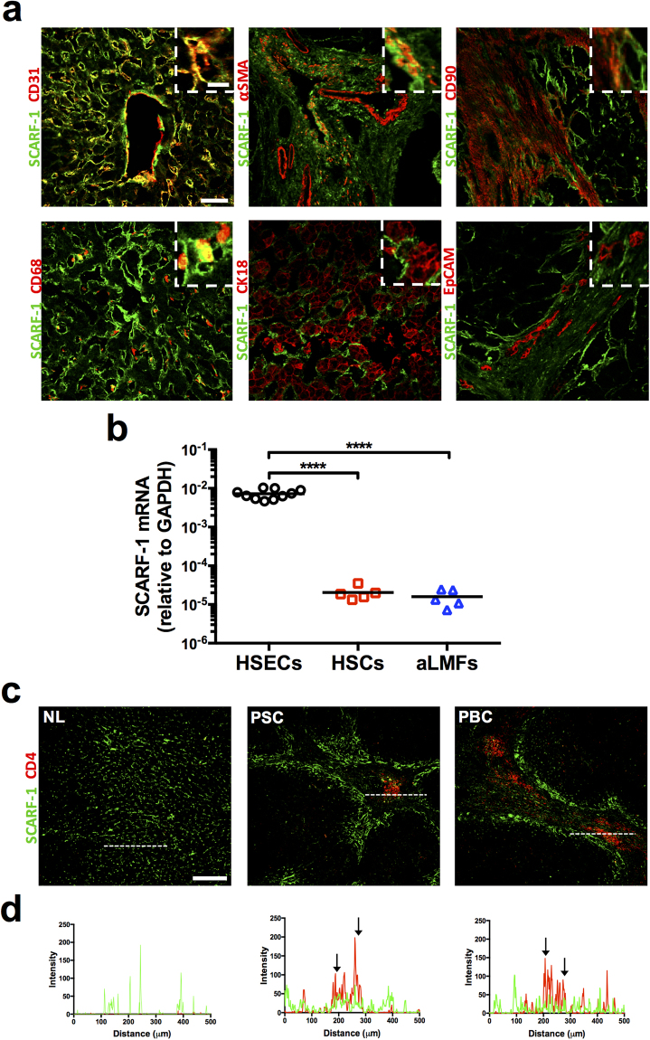
Figure 2.
HSEC represent a major cell type expressing SCARF-1 in CLD. (a) Representative images of dual colour immunofluorescent staining on chronically diseased (ALD and PSC) liver for SCARF-1 (green) and endothelial marker CD31 (top left panel), activated stellate cell marker α-SMA (smooth muscle actin; top middle panel), fibroblast marker CD90 (top right panel), macrophage marker CD68 (bottom left panel), hepatocyte marker CK18 (bottom middle panel) and biliary epithelial marker EpCAM (bottom right panel). Insets show magnification of SCARF-1 and cell-specific markers. Scale bar = 50 µm. Inset scale bar = 10 µm. (b) SCARF-1 mRNA expression in isolated human hepatic sinusoidal endothelial cells (HSECs), hepatic stellate cells (HSCs) and activated liver myofibroblasts (aLMFs). ****Indicates statistical significance where p ≤ 0.001. n = 5–10 in each group. (c) Representative images of dual colour immunofluorescent staining of SCARF-1 (green) and CD4 (red) in normal liver (NL) and chronically diseased livers (PSC and PBC). Scale bar = 250 µm. White dashed lines delineate sites of intensity measurements. (b) Intensity measurements of immunofluorescent staining shown in (a). Black arrows indicate areas of stronf co-localisation of SCARF-1 (green) and CD4 (red).

