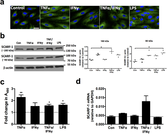
Figure 3.
In vitro expression of SCARF-1 in HSEC can be up-regulated by proinflammatory cytokines and LPS. (a) Representative images of immunofluorescent staining of SCARF-1 (green) with DAPI nuclear stain (blue). Scale bar = 25 µm. (b) Representative Western blot (left panel) and quantification (right panels) of the 180 kDa (dimeric) and 90 kDa (monomeric) species of SCARF-1 in stimulated HSEC compared to media alone control (Con). Results are representative of 3 independent experiments and are regions cropped from the same membrane (see Supplementary Figure 7). (c) Fold change in SCARF-1 protein expression measured by cell-based ELISA in stimulated HSEC. (d) qPCR analysis of SCARF-1 mRNA in stimulated HSEC. (a–d) HSEC were treated with 10 ng/ml of tumour necrosis factor (TNF)α, 10 ng/ml of interferon (IFN)γ, or both in combination or with 1 µg/ml of LPS for 24 h. (c and d) Dotted lines indicate control level of expression. * and ** indicate statistical significance where p ≤ 0.05 and p ≤ 0.01, respectively. (b and d) n = 3 and (c) n = 5 independent experiments with different HSEC donors in each group.

