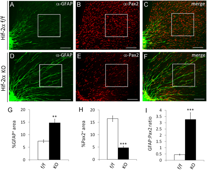
Figure 10.
Accelerated retinal astrocytic differentiation in APC specific Hif-2α knockout mice. Retinas were isolated from Hif-2α flox/flox (f/f) and Hif-2α flox/flox/GFAPCre (KO) mice at P1. After whole-mount staining with anti-Pax2 and anti-GFAP and fluorophore conjugated secondary antibodies, retinas were flat mounted and imaged by confocal microscopy. Boxed areas were quantified for astrocytic differentiation, and data are shown in (G,H, and I). n = 5 mice. **p < 0.01, ***p < 0.001. Error bars represent SEM. Scale bars are 100 µm.

