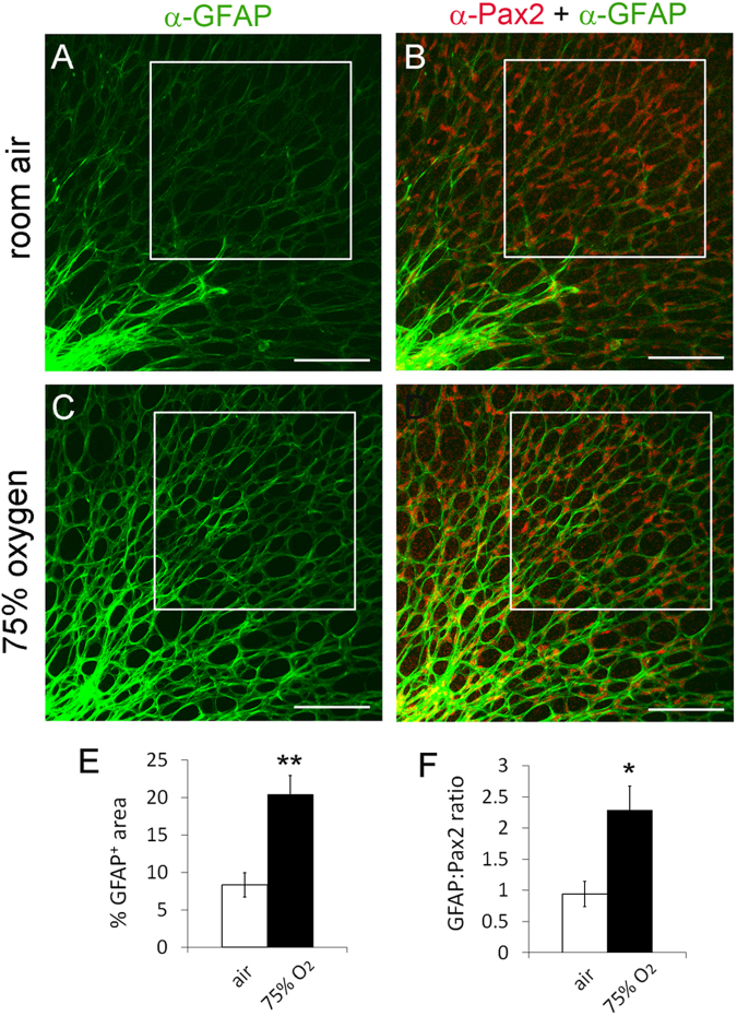
Figure 9.
Acceleration of retinal astrocytic differentiation by oxygen. At P0, wild-type CD1 mice were either maintained under room air (A) or placed into an oxygen chamber (75% O2) (B). After 24 hours, mice were euthanized, and retinas were dissected, fixed, and stained as whole-mounts by anti-Pax2 and anti-GFAP and fluorophore conjugated secondary antibodies. Stained retinas were flat-mounted and imaged by confocal microscopy. Astrocyte differentiation in boxed areas was quantified, and results are summarized in (E and F). n = 6 mice. *p < 0.05, **p < 0.01. Error bars are SEM. Scale bars are 100 µm.

