Fig. 3.
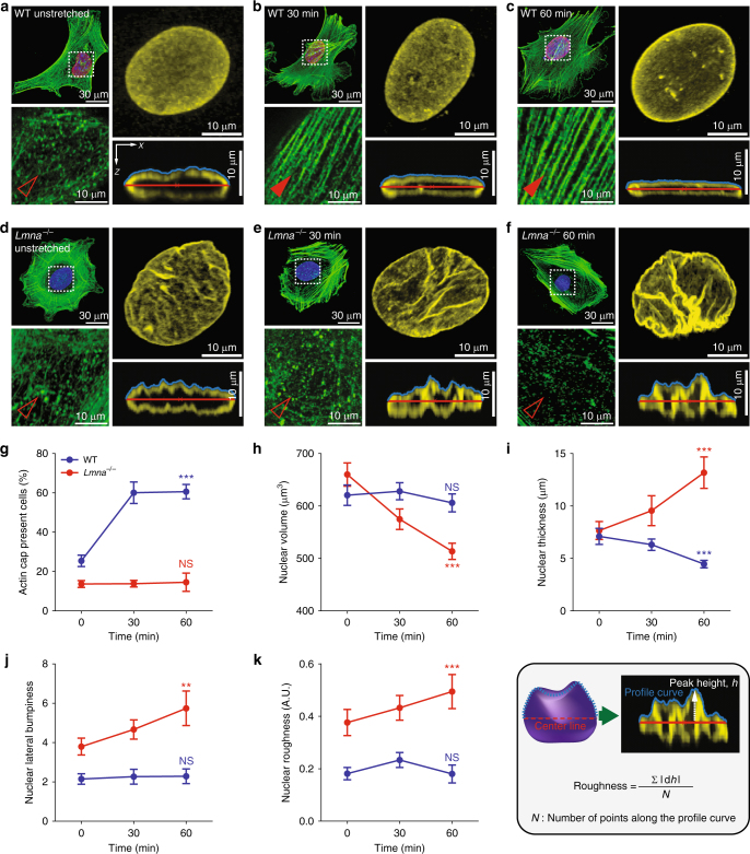
Lamin A/C-dependent differential formation of an actin cap and nuclear deformation in response to substrate stretching. a–f Representative F-actin organization and nuclear morphology of lamin A/C-present WT (a–c) and lamin A/C-deficient Lmna−/− (d–f) MEFs at different time points of the substrate stretching (0, 30, 60 min). Insets display details of F-actin organization in the apical region of the nucleus. Full and empty arrowheads indicate the presence and absence of the perinuclear actin cap, respectively. Nuclear morphology of lamin B1-stained nuclei (yellow) indicates distinct evolution of 3D-nuclear shape in response to substrate stretching, where maximum intensity projection onto the XY-plane was performed using upper hemispheres of the 3D-reconstructed nuclei to highlight the detailed nuclear surface texture. The cross-sectional side view was captured along the XZ-plane crossing the center of the nucleus. g–k Stretch-dependent formation of an actin cap (g) and changes of nuclear volume (h), thickness (i), lateral bumpiness (j), and surface roughness (k) in WT (blue) and Lmna−/− (red) MEFs. Note that nuclear flattening with volume conversation (h, i) and nuclear surface roughening with volume reduction (j, k) are the stretch-induced characteristic features of the actin cap forming WT and actin cap non-forming Lmna−/− cells (g). In i and k, nuclear thickness was defined as the maximum height in the vertically cross-sectioned image of the 3D-reconstructed lamin B1-stained nucleus and the height variation along the apical surface was termed as surface roughness. In g, >150 cells were examined per condition and in h–k, >30 lamin B1-stained nuclei were analyzed per condition. Error bars represent the S.E.M. of averaged values. Unpaired t-test was applied to compare unstretched control cells (0 min) and fully stretched cells (60 min). ***p < 0.0001, **p < 0.001, NS not significant (p > 0.05)

