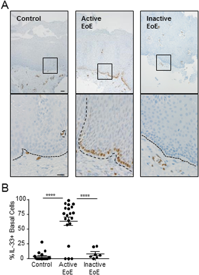
Figure 1.
Subcellular localization of IL-33 in esophageal epithelial cells. (A,B) Immunohistochemistry for IL-33 protein expression in representative esophageal biopsies from healthy control individuals (Control, left panel), patients with active EoE (Active EoE, middle panel), or patients with inactive EoE (Inactive EoE, right panel) using mouse anti–IL-33 antibody. In (A), the bottom row is a high-power view of the area enclosed in the black square. The black dashed lines indicate the basement membrane. Scale bars are both 20 µm. Biopsies from 19 controls, 20 patients with active EoE, and 7 patients with inactive EoE were stained. (B) Quantification of the proportion of basal layer cells in each biopsy with IL-33 expression. Mean ± standard error of the mean is depicted. ****p < 0.0001.

