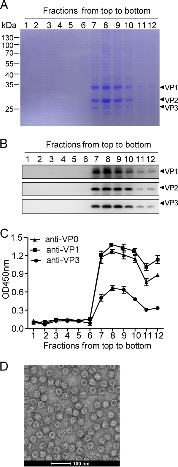
FIG 2.

Assembly of VLPΔVP4. Cell lysate from IEXBac-P1ΔVP4- and IEXBac-3CD-coinfected Sf9 cells was layered onto 10 to 50% sucrose gradients for centrifugation. Twelve fractions were taken from top to bottom and then subjected to SDS-PAGE with Coomassie blue R-250 staining (A), Western blotting (B), or ELISA with antibodies as indicated (C). The OD450 values are means and standard deviations (SD) of three triplicate wells. (D) Electron microscopy of VLPΔVP4.
