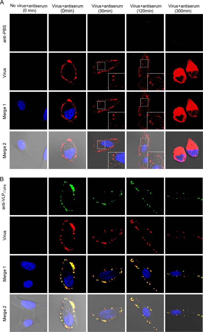
FIG 9.
Anti-VLPΔVP4 sera inhibit EV71 internalization. Vero cells were preattached to EV71 and then treated with anti-PBS (A) or anti-VLPΔVP4 (B) serum at 4°C for 1 h. After incubation at 37°C for 0, 30, 120, or 300 min, as indicated, the cells were fixed for immunofluorescence staining. Localization of virus and antibodies was visualized by confocal microscopy. The green signal represents EV71, which was detected by rabbit-anti-EV71 VLP serum; the red signal represents antibodies in anti-VLPΔVP4 serum; the blue signal represents nuclei stained by DAPI (4′,6-diamidino-2-phenylindole). Merge 1 represents a merge of green, red, and blue channels. Merge 2 represents a merge of green, red, blue, and white field channels. The insets are enlarged views of the boxed areas.

