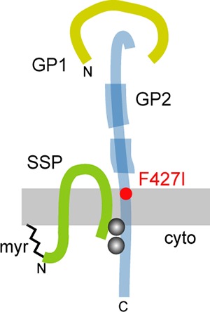
FIG 1.

Arenavirus GPC. Schematic drawing of GPC subunit organization. Features include SSP myristoylation (myr), the binuclear SSP-GP2 zinc finger (gray balls), and the N- and C-terminal heptad-repeat regions in GP2 (thick blue), diagnostic of class I viral fusion proteins. The attenuating F427I mutation in Candid#1 is shown in red. The schematic drawing is not to scale, and the relative positions of the subunits are unknown.
