
Figure 3.
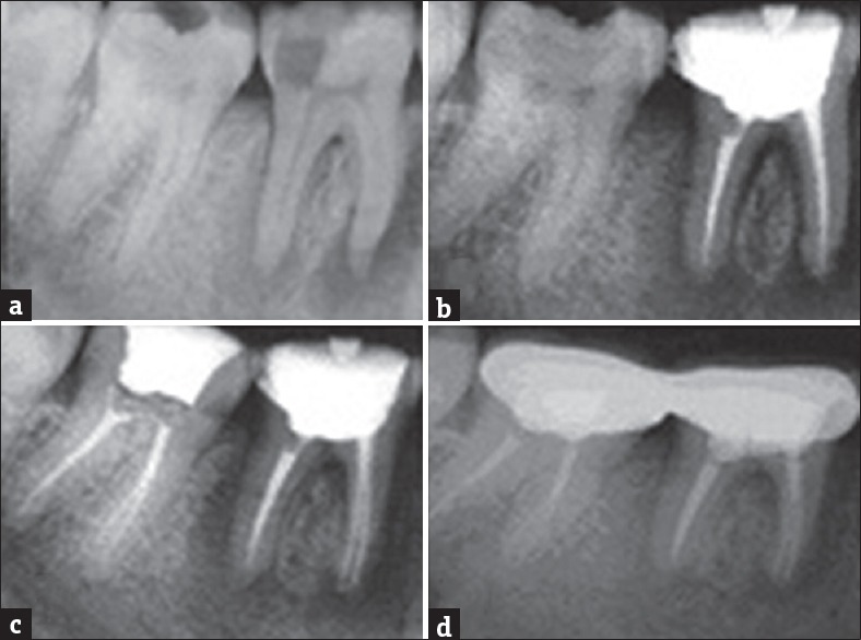
(a) Preoperative radiograph showing periapical lesion involving both roots of 36, 37. (b) Immediate postobturation radiograph 36. (c) Immediate postobturation radiograph 37. (d) One-year review showing formation of new bone
Official websites use .gov
A
.gov website belongs to an official
government organization in the United States.
Secure .gov websites use HTTPS
A lock (
) or https:// means you've safely
connected to the .gov website. Share sensitive
information only on official, secure websites.
(a) Preoperative radiograph showing periapical lesion involving both roots of 36, 37. (b) Immediate postobturation radiograph 36. (c) Immediate postobturation radiograph 37. (d) One-year review showing formation of new bone