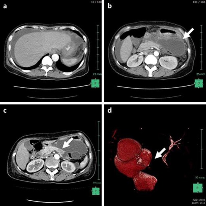
Fig. 1.
a, b A contrast-enhanced computed tomography (CT) of the abdomen on admission day revealed massive fluid collection in the abdominal cavity and gigantic multilocular cysts of the pancreatic tail (white arrow). c A contrast-enhanced CT after recovery from acute peritonitis revealed the 2-cm low-density mass located at the pancreas body (white arrow) and the peripheral main pancreatic duct (MPD) dilatation. d A magnetic resonance cholangiopancreatography also showed interruption of the MPD (white arrow) and dilatation of the peripheral MPD dilatation.

