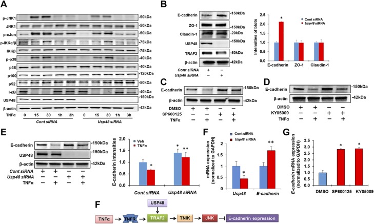
Figure 6.
Knockdown of USP48 attenuates TRAF2-dependent activation of JNK1, therefore increasing E-cadherin expression. A) Knockdown of USP48 attenuates TNF-α-induced phosphorylation of JNK1 and c-Jun. Beas2B cells were transfected with control siRNA or Usp48 siRNA, and incubated for 3 d. Cells were then treated with TNF-α (10 ng/ml) for 0 to 3 h. Cell lysates were examined by immunoblotting with indicated antibodies. B) Knockdown of USP48 increases E-cadherin expression. Beas2B cells were transfected with control siRNA or Usp48 siRNA for 3 d. Then E-cadherin, ZO-1, claudin 1, USP48, TRAF2, and β-actin levels were analyzed by Western blot analysis (n = 3). *P < 0.01 compared to control siRNA transfected cells. C) Inhibition of JNK attenuates TNF-α-induced reduction of E-cadherin. Beas2B cells were pretreated with SP600125 for 1 h. Cells were then added with TNF-α (10 ng/ml) for 48 h. E-cadherin and β-actin levels were analyzed by Western blot analysis. D) Inhibition of TNIK attenuates TNF-α-induced reduction of E-cadherin. Beas2B cells were pretreated with KY-05009 for 1 h. Cells were then added with TNF-α (10 ng/ml) for 48 h. E-cadherin and β-actin levels were analyzed by Western blot analysis. E) Knockdown of USP48 attenuates TNF-α-induced reduction of E-cadherin. Beas2B cells were transfected with control siRNA or Usp48 siRNA and incubated for 72 h. Cells were then treated with TNF-α (10 ng/ml) for 48 h. E-cadherin, USP48, and β-actin levels were analyzed by Western blot analysis (n = 3). *P < 0.01 compared to vehicle + control siRNA; **P < 0.01 compared to TNF-α + control siRNA. Shown are representative blots from at least 3 independent experiments. F) Knockdown of USP48 increases E-cadherin mRNA levels. Beas2B cells were transfected with control siRNA or Usp48 siRNA and incubated for 3 d. RNA was extracted and analyzed by real-time qPCR with Usp48 and E-cadherin primers. Relative expression of Usp48 and E-cadherin was normalized to Gapdh (n = 3). *P < 0.01 compared to control siRNA; **P < 0.01 compared to control siRNA. G) Inhibition of JNK or TNIK increases E-cadherin mRNA levels. Beas2B cells were treated with SP600125 (10 μM) or KY-05009 (10 μM) for 24 h. RNA was extracted and analyzed by real-time qPCR with E-cadherin primers. Relative expression of E-cadherin was normalized to Gapdh. *P < 0.01 compared to DMSO. H) Scheme showing down-regulation of USP48 or inhibition of TNIK and JNK increases E-cadherin expression. Data are presented as means ± sd.

