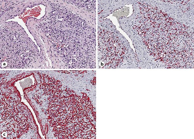
Fig. 3.
a Dense proliferation of endothelial cells without nuclear atypia, forming capillary vessels with barely identifiable vascular lumina, surrounding a normal vessel. b Nuclear expression of ERG, a highly specific marker for vascular endothelium. c Newly formed capillary vessels show a normal anatomic architecture of blood vessels with an outer layer of α-SMA-positive pericytes. a Hematoxylin-eosin stain. Original magnification: a–c ×200.

