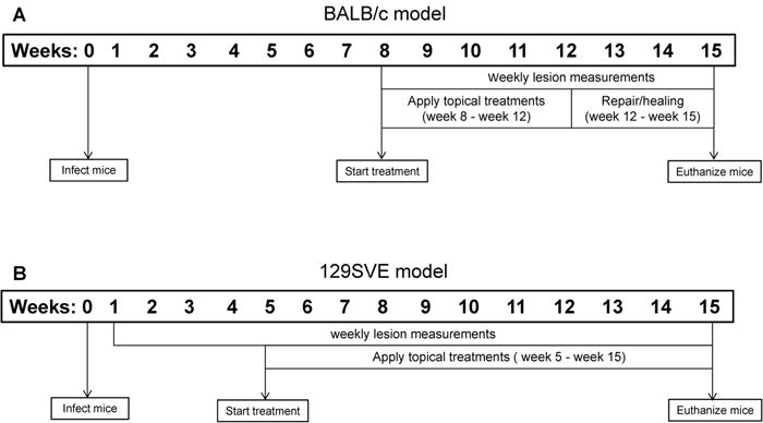
Fig. 1.

Timeline of study. (A) For the ulcerated model, BALB/c mice were infected with L. mexicana promastigotes subcutaneously at their back rumps. After establishment of ulcerated lesions (week 8 post infection) mice were randomized into two groups and treated for 4 weeks. Serum was collected at weeks 8, 12 and 15. Mice were euthanized at week 15 and lesions and lymph node cells were collected for analysis. (B) For the non-ulcerated model, 129SVE mice were infected with L. mexicana promastigotes subcutaneously at their back rumps. After development of visible lesions (week 5 post infection) mice were randomized into two groups treatments were applied for 10 weeks (weeks 5–15 post infection). Serum was collected at weeks 8, 11 and 15. Mice were euthanized at week 15 and lesions and lymph node cells were collected for analysis.
