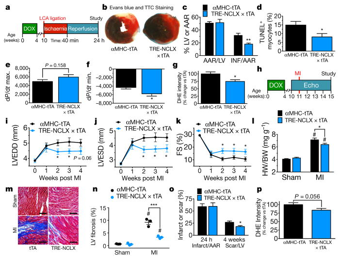
Figure 4. Overexpression of NCLX protects against myocardial ischaemic reperfusion injury and ischaemic heart failure.
a, Timeline of myocardial ischaemia reperfusion experimental protocol. DOX, doxycycline administration. b, Mid-ventricular cross-sections of hearts 24 h after reperfusion and EBD perfusion and TTC staining (non-blue area indicates the area at risk (AAR); red area, viable myocardium; white area, infarct). c, Per cent AAR of the left ventricle and per cent infarct of the AAR. n = 11 mice per group. d, TUNEL+ cardiomyocytes post ischaemia reperfusion. n = 5 mice per group. e, f, Invasive haemodynamics post ischaemia reperfusion. e, dP/dt maximum (contractility). f, dP/dt minimum (relaxation). n =13–16 mice per group. g, DHE staining in live myocardium 24 h post ischaemia reperfusion. n = 6 tTA and n =5 NCLX-Tg. h, Timeline of myocardial infarction (MI) experimental protocol. Echo, left ventricular echocardiography. i, Left-ventricular end-diastolic dimension (LVEDD). j, Left-ventricular end-systolic dimension (LVESD). k, Per cent fractional shortening (FS). n =19 tTA, n =16 NCLX-Tg. l, Heart weight to body weight ratio. n = 6, sham tTA, n = 7 sham NCLX-Tg, n =17 tTA, n =17 NCLX-Tg. m, Images of Masson’s trichrome-stained left ventricular sections at the border zone 4 weeks post myocardial infarction. Scale bars, 400 μm. n, Per cent fibrosis in peri-infarct and remote regions of the left ventricle 4 weeks post infarction. n = 3 mice per group. o, Per cent infarct of AAR 24 h after LCA ligation and scar of left ventricle 4 weeks post infarction. n = 5 mice per group. p, DHE staining in live myocardium 4 weeks post infarction. n = 8–9 mice per group. *P <0.05, **P <0.01, ***P <0.001; #P < 0.05 versus sham control (l, n).

