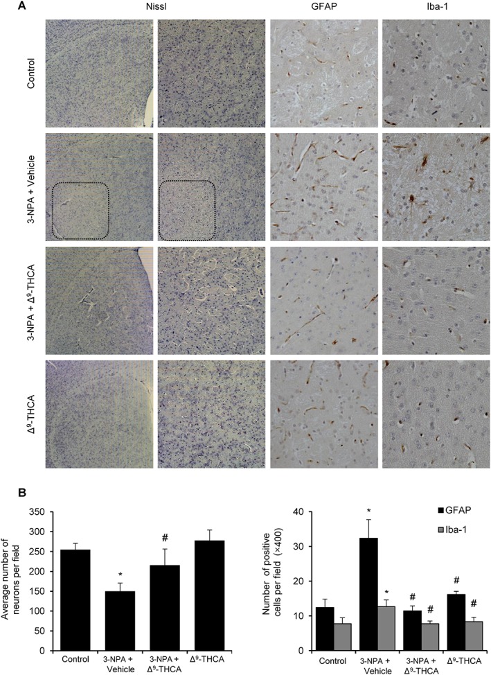
Figure 7.

Δ9‐THCA prevents neuronal loss, microgliosis and astrogliosis induced by 3‐NPA administration. (A) Representative images of Nissl, Iba‐1 and GAFP staining performed on coronal striatal brain sections (original magnification 20×). (B) Quantification of the different markers was performed with ImageJ software. Total average number of neurons (Nissl), microglia (Iba‐1+) and astrocytes (GFAP+) is shown. Values are expressed as means ± SEM (n = 6). * P < 0.05, significantly different from control group; # P < 0.05, significantly different from 3‐NPA group (n = 9 animals per group).
