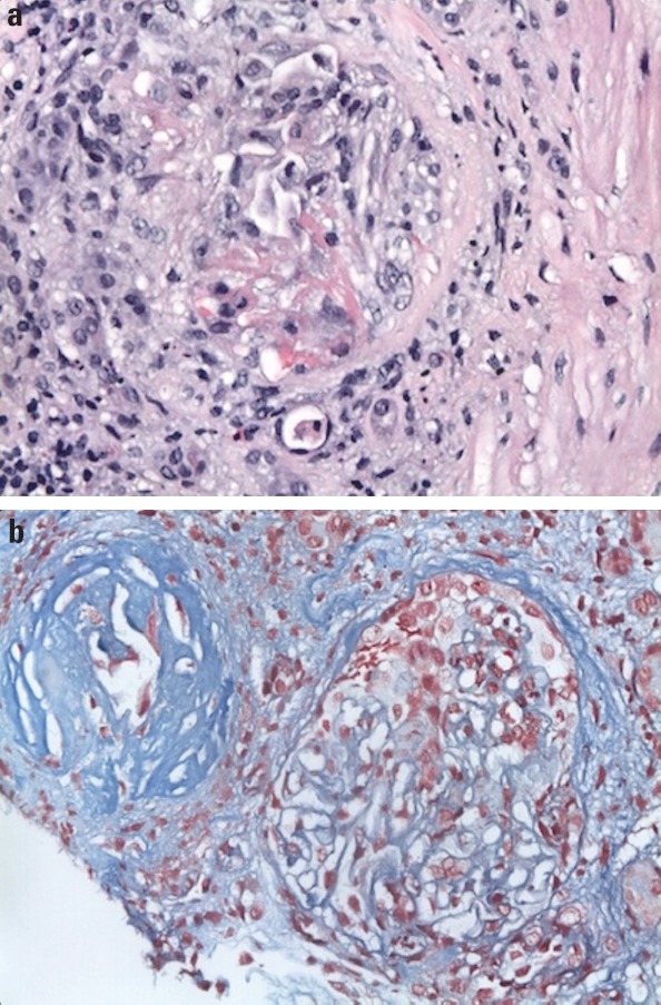
Figure 3.

Renal biopsy demonstrates (a) sclerotic glomeruli with segmental cellular crescent formation in a background of mixed interstitial inflammation composed of mature lymphocytes, plasma cells, and eosinophils (400× hematoxylin and eosin stain) and (b) shrunken globally sclerotic glomeruli and fibrocellular crescent glomerulus with fibrin deposition-red on trichrome stain (400× trichrome)
