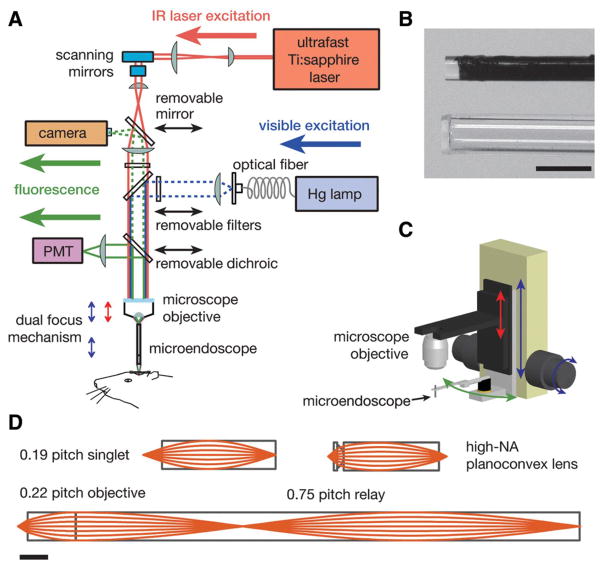
FIGURE 1.
Methodologies for in vivo optical microendoscopy. (A) Optical schematic of an upright microscope modified to permit both one- and two-photon fluorescence microendoscopy. For two-photon imaging, the beam from an ultrashort-pulsed infrared (IR) Ti:sapphire laser is scanned within the focal plane of the microscope objective. By adjusting the axial separation between the objective and the microendoscope (red arrow of the dual-focus mechanism; see also C), this focal plane of the microscope objective is also set to the microendoscope’s back focal plane. Another focal adjustment (blue arrows of the dual mechanism) is used to lower the objective and the microendoscope in tandem toward the animal. For one-photon imaging, a mercury (Hg) arc lamp provides illumination. In both imaging modes, fluorescence emissions route back through the microendoscope and to either a camera or a photomultiplier tube (PMT) for one- or two-photon imaging, respectively. (B) Photographs of the tips of a 0.5-mm-diameter micro-endoscope of doublet design (top) and a 0.8-mm-outer-diameter glass capillary guide tube (bottom) into which this microendoscope can be inserted. The relay of the microendoscope is coated black. A glass coverslip is attached to the tip of the guide tube. The guide tube facilitates the rapid exchange of microendoscopes without perturbation to the underlying tissue. Scale bar, 1 mm. (C) The microscope objective and the microendoscope probe are mounted on a pair of cascaded focusing actuators that provide dual-focus capability. This allows the objective to be moved either alone (red arrow) or together with the microendoscope (blue arrow). The microendoscope can also be swung out of the optical axis (green arrow) to permit conventional microscopy. (D) Optical ray diagrams for sample microendoscopes of the singlet GRIN (top left), compound plano-convex and GRIN (top right), and GRIN doublet (bottom) types. Scale bar, 1 mm.

