
Figure 1. Optics and Protein Engineering Converge for Ca2+ Imaging in >1,200 CA1 Pyramidal Cells in Freely Moving Mice.
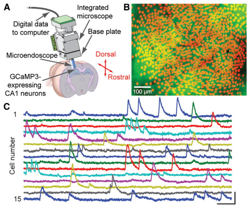
(A) An integrated microscope is equipped with a microendoscope to image CA1 neurons expressing the engineered Ca2+ indicator GCaMP3 under control of the Camk2a promoter. The base plate and microendoscope are fixed to the cranium for repeated access to the same field of view. Republished from Ziv et al., 2013.
(B) Shown are 1,202 CA1 pyramidal cells (red somata) identified by Ca2+ imaging in a freely moving mouse atop a mean fluorescence image (green) of CA1. Vessels appear as dark shadows. Image courtesy of Yaniv Ziv and Lacey Kitch, Stanford University.
(C) Example traces of [Ca2+]i dynamics from 15 cells. Scale bars denote 5% ΔF/F (vertical) and 10 s (horizontal).