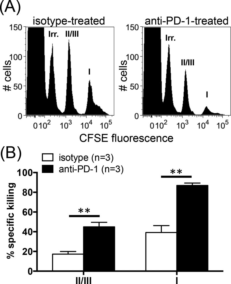
Fig. 4.
Anti-PD-1 treatment augments subdominant TCD8-mediated cytotoxicity. Target cells were prepared by pulsing syngeneic naïve splenocytes with gB498 (irrelevant peptide), site II/III peptide or site I peptide, which were labeled with 0.025 µM, 0.25 µM and 2 µM CFSE, respectively. Target cells were mixed in equal numbers and injected into the tail vein of C57SV-primed mice that had received anti-PD-1 or isotype control (n=3 per group). Four h later, target cell populations were tracked by flow cytometry in each spleen (A), and their % specific lysis was calculated (B). ** denotes a statistical difference with p<0.01 by unpaired Student’s t-test. Error bars (B) represent SEM.

