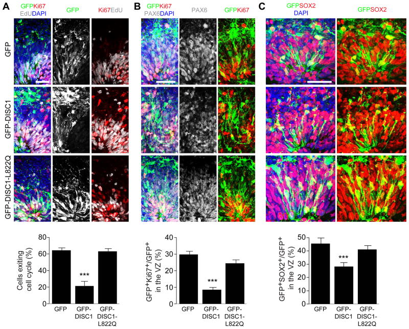
Figure 5. Interaction between DISC1 and Ndel1 regulates the cell cycle of RGCs in human forebrain-specific organoids.
(A) Expression of DISC1 765–835 peptide leads to delayed cell cycle progression of RGCs in human forebrain organoids. Human forebrain organoids generated from human iPSC line C3-1 were electroporated with GFP (GFP), GFP-DISC1 765–835 (GFP-DISC1) or GFP-DISC1 765–835 L822Q (GFP-DISC1-L822Q) plasmids at day 45–47 in culture, and further cultured for 5 days before analysis. Shown are representative sample confocal images of organoids under different conditions pulsed with EdU for 2 hr at 4 days after electroporation and immunostained with GFP, Ki67, EdU and DAPI. Cell exit index is calculated by GFP+EdU+Ki67-/GFP+EdU+ cells.
(B) Effect of DISC1 765–835 peptide expression on NSC proliferation. Shown are representative sample confocal images of organoids under different conditions immunostained with GFP, Ki67, PAX6 and DAPI. Quantification of proliferating RGCs (Ki67+) is shown at the bottom.
(C) Effect of DISC1 765–835 peptide expression on the maintenance of the RGC population. Shown are representative sample confocal images of organoids under different conditions immunostained with GFP, SOX2 and DAPI. Quantification of total SOX2+ RGCs is shown at the bottom. Scale bars: 50 μm. Numbers associated with each bar graph refer to the total number of neural tube structures analyzed under each condition. Values represent mean ± SEM (***P < 0.001, One-way ANOVA).
See also Figure S6.

