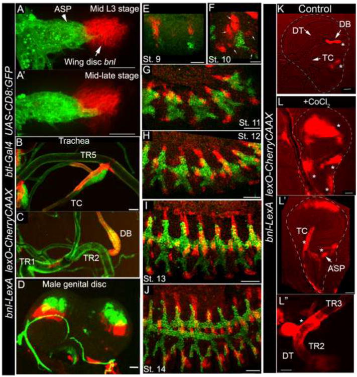
Figure 3. Expression of bnl-LexA in developmental stages and in hypoxia. A-A′.

Wing disc bnl source (red) and the ASP (green) in mid and late L3 larval wing discs. B-C; bnl expression in larval tracheal branches: TR5 transverse connective (TC) (B); TR2 dorsal branch (DB) (C). D, bnl expression (red) in genital discs. Mesenchymal cells associated with the bnl expressing disc epithelium express btl (green). E-J, confocal images of fixed embryos of different stages; Small arrows, five bnl sources surrounding the tracheal placode. Genotypes: A-J; btl-Gal4, UAS-CD8:GFP/+; bnl-LexA,lexO-herryCAAX/+. K-Lhypoxia induced bnl-LexA expression profile in wing discs and associated TR2 tracheal metamere (TC, DB, dorsal trunk-DT). Genotype: bnl-LexAlexO-CherryCAAX/+. K, control discs from ex vivo cultured organs without CoCl2. L-Lwing discs (L, L′) and trachea (DT, (L″)) from cultured ex vivo organs with CoCl2 induced hypoxia. Star, ectopic expression. K-L″; Images were captured with 555 nm fluorescence as well as with bright field. The superimposed images were shown. Scale bars, 30 μm; K-L″, 50 μm.
