
Figure 6. Neuronal expression of bnl in larval and embryonic development. A.
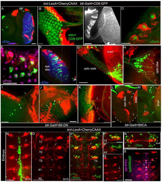
bnl-LexA and btl-Gal4 expression in larval eye disc; Elav immunostaining (blue) marked retinal neurons posterior to the morphogenetic furrow (MF). B, bnl-LexA expression marked neurons in ommatidial clusters; elav-Gal4 driven CD8:GFP marked all neurons (UAS-CD8:GFP X elav-Gal4). C, ommatidial bnl-neuron extended axons through optic stalk into the optic lobe. D, 3D projection showing bnl-expressing neurons marked by Sens immunostaining, a marker for the R8 photoreceptors. E, 3D projection showing Pros immunostaining that marked btl-expressing neurons; Pros, R7 photoreceptor marker. F, 3-D projection showing intimate organization of btl- (green) and bnl- (red) expressing neurons in the eye disc ommatidia (also see supplementary movies 12,13); F-H, axons from receptor (Btl) and ligand (Bnl) expressing neurons fasciculate through optic stalk into the optic lobe; arrow, fasciculation in optic lobe. I-M, phenotypic consequences in bnl- and btl- expressing neurons when Btl-DN (I-K) and λBtl (L-M) were expressed specifically in the btl-neurons; arrow, bnl-expressing neurons; arrowhead, btl-expressing neurons; star, hemocytes; dashed line (L-M), eye disc out line; L, M, same sample in two different magnifications. Genotypes: A, E- H, btl-Gal4, UAS-CD8:GFP/+; bnl-LexA,lexO-CherryCAAX/+; C, D, bnl-LexAlexO-CherryCAAX/+; I-K, btl-Gal4, UAS-CD8:GFP/+; bnl-LexA,lexO-CherryCAAX/UAS-Btl-DN; L, M, btl-Gal4, UAS-CD8:GFP/UAS-XBtl; bnl-LexA, lexO-CherryCAAX/+. A-H, eye disc anterior-left. N-S, stage 14-16 embryos, ventral view, anterior up. N-O, bnl-LexA expression in neurons (red) and btl-Gal4 (N) in the ventral midline (green) of stage 14 (N) and stage 15 (O) embryos; bnl-LexA expressing neurons in thoracic segments (T1-T3), had both interneuronal (arrowhead) and motor neuronal (arrow) projections, and had only interneuronal projections in abdominal hemisegments. P-P″, immunostaining for Ems, a marker for NB 3-5 and 4-4 lineages, marked bnl-expressing neurons in T1-T3, but did not mark the abdominal ones; P′,P″, magnified view of Ems immunostaining from T3 (P) and A2 clusters (P″) shown in P. Q; punctate lobular fluorescence (arrow) next to the large cells bodies of the abdominal bnl-expressing NB lineage suggested apoptosis in several abdominal neurons. R, one cell (arrow) in each bnl-cluster is immunostained for Dpn, a neuroblast marker. S, synaptic contacts (green fluorescence) between btl-expressing cells (purple) and bnl-neurons (red), mapped (arrow) in the ventral midline by GFP reconstitution using nsyb-GRASP technique; genotype: btl-Gal4/ lexO-nsybGFP1-10, UAS-CD4:GFP11; bnl-lexA, lexO-CherryCAAX/UAS-CD4:IFP2. Scale bars, 30 μm; A, 60 μm; D, 10 μm.