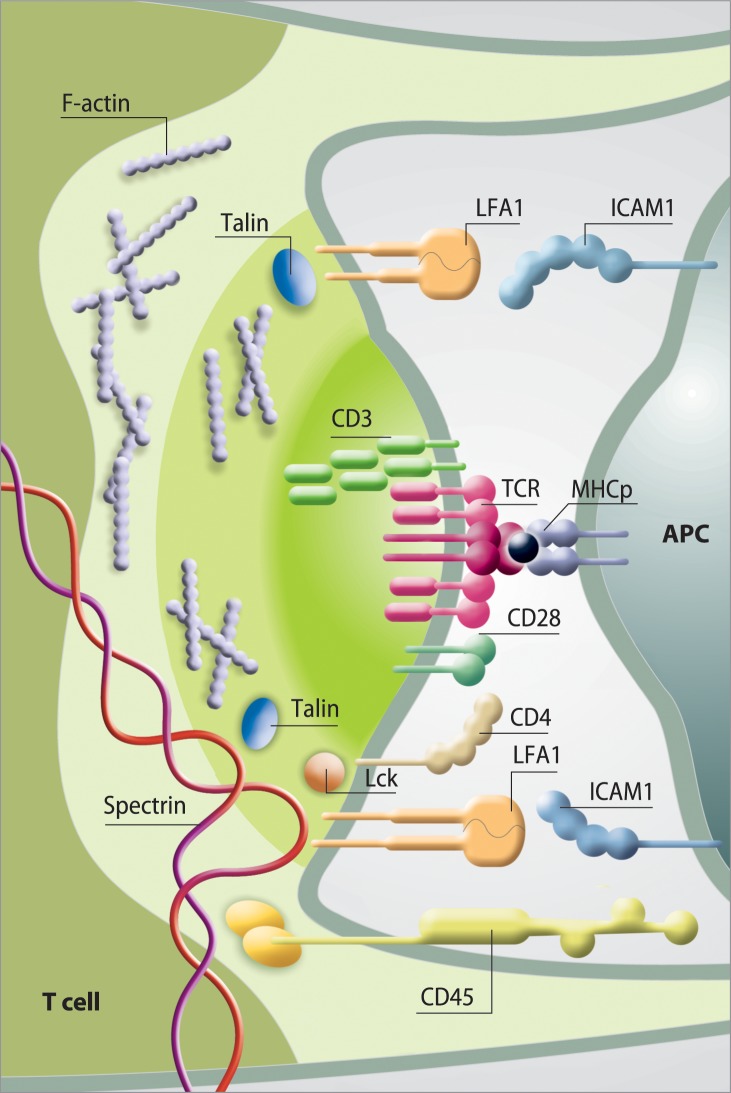
Fig 1. Schematic of the immunological synapse (IS) and representative protein interactions in the synaptic space.
Distribution of receptors and adhesion molecules in individual clusters in the immune synapse. The T-cell receptor (TCR) / CD3 complex interacts with MHC-peptide. The adhesion molecules on the surface of both cells (LFA-1—ICAM- 1 are responsible for the formation and stabilization of the IS, as well as for initiating signal transduction pathways activated by TCR. The distal ring of IS is rich in proteins, such as CD45 and F-actin controls lamellipodia and filopodia formation.

