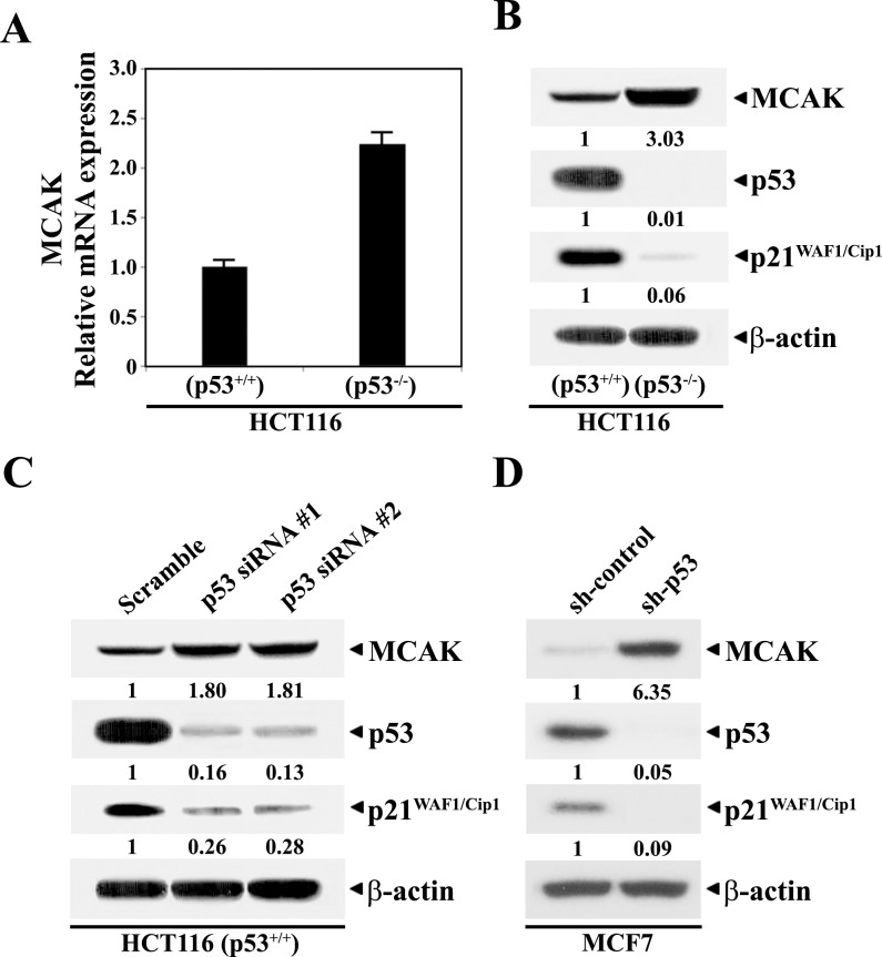
Fig 1.
Semi-quantitative RT-PCR analysis of mRNAs specific for MCAK and GAPDH (A), western analysis of protein levels of MCAK, p53, p21WAF1/Cip1, and β-actin in continuously growing HCT116 (p53+/+) and HCT116 (p53−/−) cells (B), and western blot analysis of individual protein levels in HCT116 (p53+/+) cells following treatment with the control scramble RNAi or Stealth RNAi for p53 (C), and in MCF7 cells stably transfected with control shRNA vector or shp53 vector (D). RT-PCR was performed using gene-specific primers as described in Materials and methods. To ensure that the same amount of RNA was being used, the total RNA concentration was normalized to that of GAPDH as the message of a housekeeping gene. HCT116 (p53+/+) cells were treated with each RNAi for 48 h. Western blot analysis was performed using ECL Western Blotting Kit as described in Section 2. A representative study is shown and two additional experiments yielded similar results.

