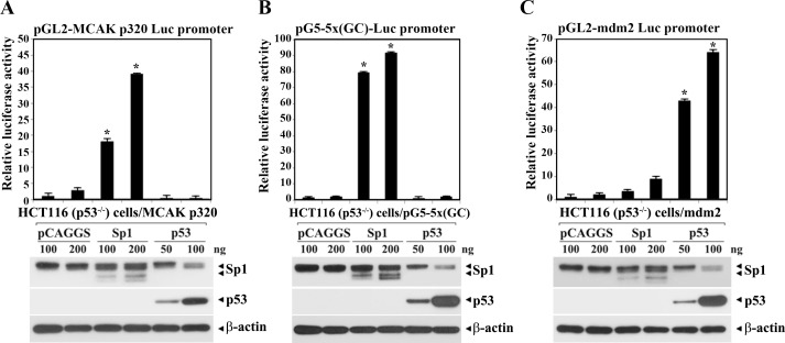
Fig 5.
Differential effect of Sp1 and p53 on the MCAK core promoter (pGL2-320-Luc) activity (A), the pG5-5×(GC)-Luc activity (B), and pGL2-mdm2 promoter (pGL-mdm2-Luc) activity (C). One hundred nanograms of individual promoter-reporter constructs (pGL2-320-Luc, pG5-5×(GC)-Luc, and pGL2-mdm2-Luc) were cotransfected with pCAGGS-Sp1, pCAGGS-wt-p53, or pCAGGS control empty vector into HCT116 (p53-/-) cells, and then the luciferase activity was determined. Each value is expressed as the mean ± SD (n = 3). *Statistical significance was defined as p<0.05 compared to the control (100 ng of pCAGGS). To analyze the expression level of MCAK, Sp1, and β-actin proteins in the cells after transient transfection, western blot analyses were performed as described in Materials and methods. A representative study is shown and two additional experiments yielded similar results.

