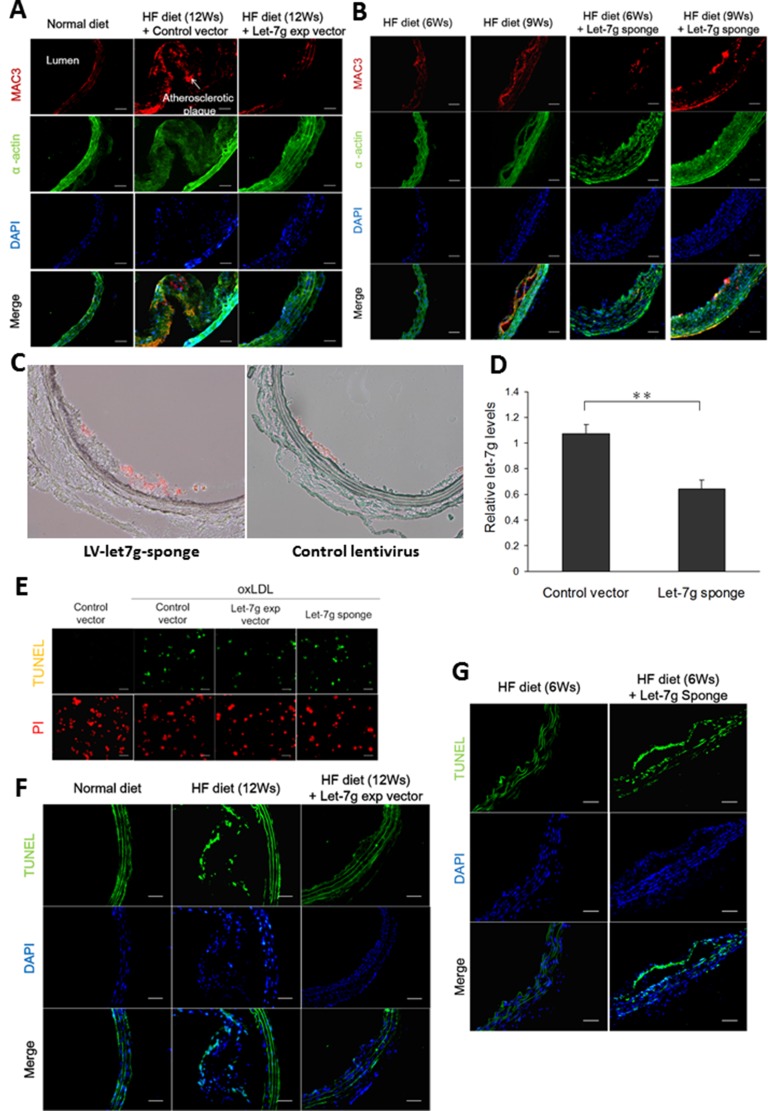
Figure 2. Let-7g decreases macrophage accumulation in the plaques, reduces the plaque size and exerts anti-apoptotic effect.
(A) Atherosclerotic plaques developed in the aortas of apoE KO mice (n = 6 per group) under a HF diet for 12 weeks, but the plaque sizes were substantially reduced when the animals were injected with LV-let7g. Macrophages (red) and VSMC (green) were indicated by the MAC3 and α-actin immunfluorescent staining, respectively. Scale bar, 50 μm. (B) The atherosclerotic plaques of apoE KO mice (n = 6 per group) injected with LV-let7g-sponge under a HF diet for 6 or 9 weeks. Scale bar, 50 μm. (C, D) Representative aortic slices from apoE KO mice treated with LV-let7g-sponge (left) or control lentivirus (right) for 9 weeks while under a HF diet. Macrophages (red) were indicated by the MAC3. let-7g level in the macrophages dissected from the atherosclerotic plaques by laser capture micro-dissection was quantified in the bar chart. To measure let-7g levels, RNU6B was used as the internal control. (E) The TUNEL assay for macrophages infected with LV-let7g, LV-let7g-sponge or LV-control vector. Scale bar, 100μm. (F, G) TUNEL-positive cells (green) in aortic sections of apoE KO mice (n = 6 per group) treated with LV-let7g or LV-let7g-sponge. Scale bars, 50 μm. The data were from at least three independent experiments. Data in each bar chart are presented as mean ± S.E.M. *P < 0.05, **P < 0.01, ***P < 0.001.

