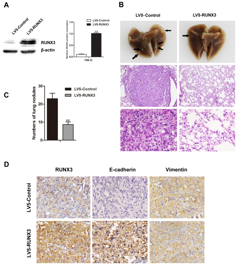
Figure 3. RUNX3 suppressed RCC metastasis in vivo.
Nude mice were injected intravenously with 786-O cells stably expressing LV5-RUNX3 or LV5-Control. Western blot was used to examine RUNX3 level A. Representative image of lung with metastatic nodules (top) and H&E staining (middle ×100, bottom ×400) 2 months after injection B. Arrows indicate metastatic nodules. The number of lung metastatic nodules was counted under a dissecting microscope C. Fewer lung metastases were seen in the LV5-RUNX3 group compared with controls. Data are shown with means ± SD (**P<0.01). Immunostaining of RUNX3, E-cadherin and vimentin in metastatic nodules of LV5-RUNX3 or LV5-Control groups D. E-cadherin expression was higher in the LV5-RUNX3 group compared with controls; vimentin was unchanged.

