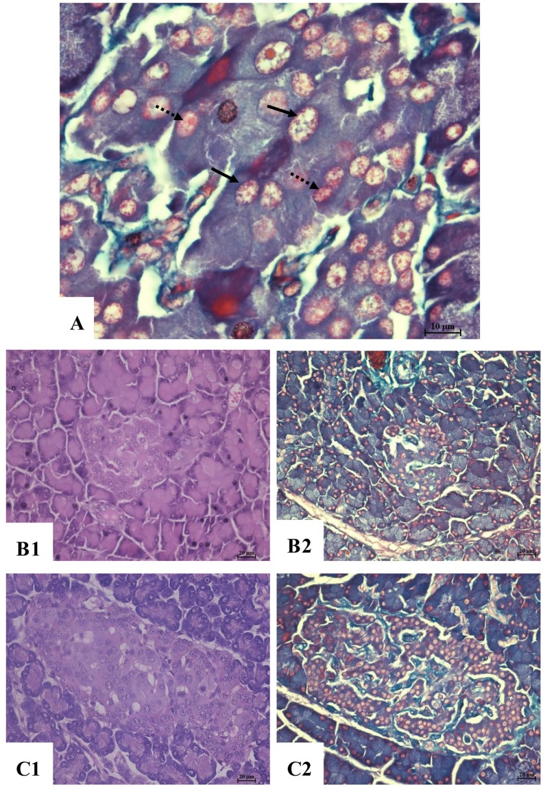
Figure 4. Histopathological changes of pancreatic islet tissue.
(A) aldehyde fuchsin staining ×1000, α cell (arrow) red orange colored granules in the cytoplasm, with a light orange colored nucleus; β cell (dot arrow), purplish red colored granules in the cytoplasm, with bright purple colored nucleus. (B1) Negative control, H&E, ×400. (B2) Negative control aldehyde fuchsin staining, ×400. (C1) High dose, H&E, ×400. (C2) High dose, aldehyde fuchsin staining, ×400.

