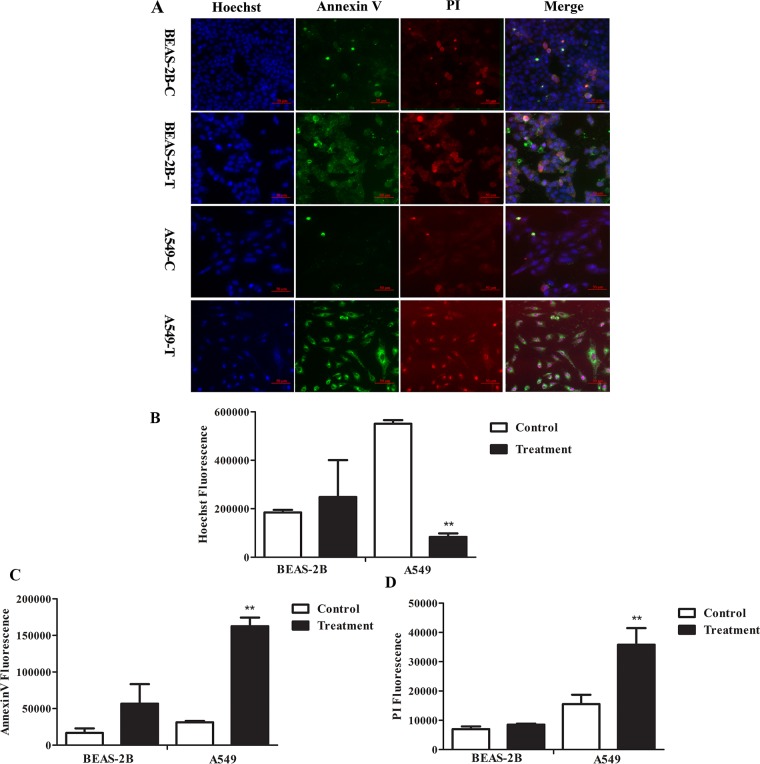
Figure 2. The effects of FMG on cell apoptosis.
Cells were treated with FMG for 48 h, and then stained with Annexin V-FITC/PI fluorescence dyes. (A) Representative images of stained cells were taken by the high-content screening (HCS) AssayScan VTI Reader (×100). Quantification of the Hoechst 33342 (B), Annexin V-FITC (C) and PI (D) fluorescent intensity by the HCS AssayScan software. Values expressed as mean ± SD from three independent experiments, *P < 0.05, **P < 0.01 vs. control group.

