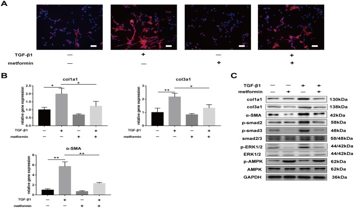
Figure 5. Metformin decreases TGF-β1-induced adhesion in vitro.
(A) Immunofluorescent (IF) staining showed that the expression of α-SMA positive (red) fibroblasts was inhibited by metformin treatment for 24 h. scale bar = 50 μm. (B) The real-time PCR analysis showed that mRNA expression levels of col1a1, col3a1 and α-SMA in NIH/3T3 fibroblasts were inhibited by metformin treatment. (C) The western blot analysis showed that protein levels of fibrotic genes were inhibited by metformin treatment. Metformin treatment increased phosphorylation of AMPK, and inhibited both SMAD and MAPK signaling pathways in NIH/3T3 fibroblasts. Data are expressed as means ± SEM. * P < 0.05; ** P < 0.01.

