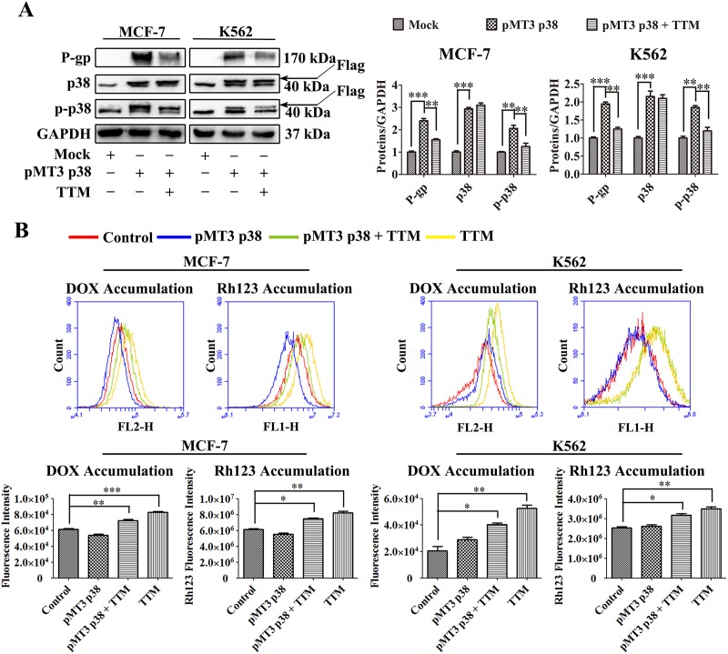
Figure 6. Effect of TTM on p38 MAPK-mediated increase in P-gp expression in parental MCF-7 and K562 cells.
(A) Representative western blot analysis of P-gp and total and phosphorylated p38 MAPK levels in control and p38 MAPK overexpressing MCF-7 and K562 cells treated with or without 30 μM TTM for 48 hr. MCF-7 and K562 cells were transfected with (+) or without (-) 10 μg/l pMT3-p38MAPK plasmid for 24 hr to increase p38 MAPK expression. GAPDH was used as a loading control. Note: ** denotes P < 0.01 and *** denotes P < 0.001 compared to the pMT3 p38 group. (B) Flow cytometry analysis of DOX and Rh123 accumulation in control and p38 MAPK overexpressing MCF-7 and K562 cells treated with or without 30 μM TTM for 48 hr. After 48 hr, the cells were incubated with 10 μM DOX for 4 hr or 5 μM Rh123 for 2 hr. Intracellular fluorescence was analyzed by flow cytometry. The data are presented as the mean ± SEM from three independent experiments. Note: * denotes P < 0.05, ** denotes P < 0.01 and *** denotes P < 0.001 compared to the control group.

