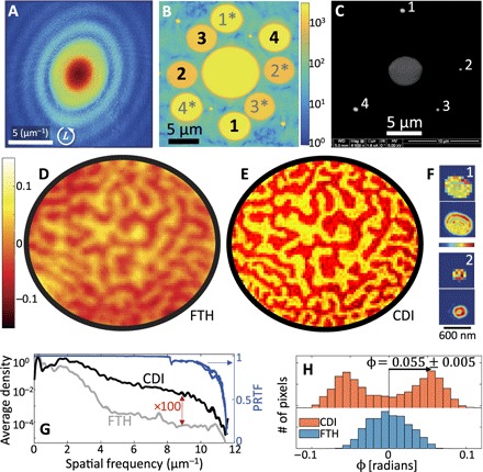
Fig. 2. Experimental results and the retrieval of nanoscale magnetic domain structure using high-order harmonics.

(A) Diffraction pattern recorded with a left-hand circularly polarized 21-nm high-harmonic beam (logarithmic color scale, 341 s total integration time, 9 × 10⁸ detected photons, corrected for spherical aberration by projection onto the Ewald sphere). (B) Fourier transform magnitude of the recorded diffraction pattern (logarithmic color scale). The holographic reconstructions from reference holes are marked 1 to 4 (conjugate holograms, 1* to 4*). (C) SEM micrograph of the gold-coated side of the sample, showing the central aperture (gray, 5 μm diameter) and four reference holes (white, numbered). (D and E) Magneto-optical phase-contrast images of the worm-like domain structure, obtained by FTH (reference hole 2, 250 nm diameter) and by CDI reconstruction, respectively (true pixel resolution, no interpolation). Positive (negative) phase indicates magnetization parallel (antiparallel) to the HHG beam. (F) Magnitude (linear scale, magnified) of the reconstructed exit waves of reference holes 1 and 2, at a true pixel resolution (top) and interpolated (bottom), illustrating the presence of high-order waveguide modes within the aperture. (G) Left axis: Fourier spectral density of phase-contrast images (azimuthally averaged). Right axis: Phase retrieval transfer functions (PRTFs) for independent reconstructions of left- and right-handed incident circular polarizations, demonstrating high fidelity of the CDI reconstructions. (H) Histogram of the measured phase, as reconstructed using CDI and FTH, allows quantitative evaluation of the magneto-optical phase.
