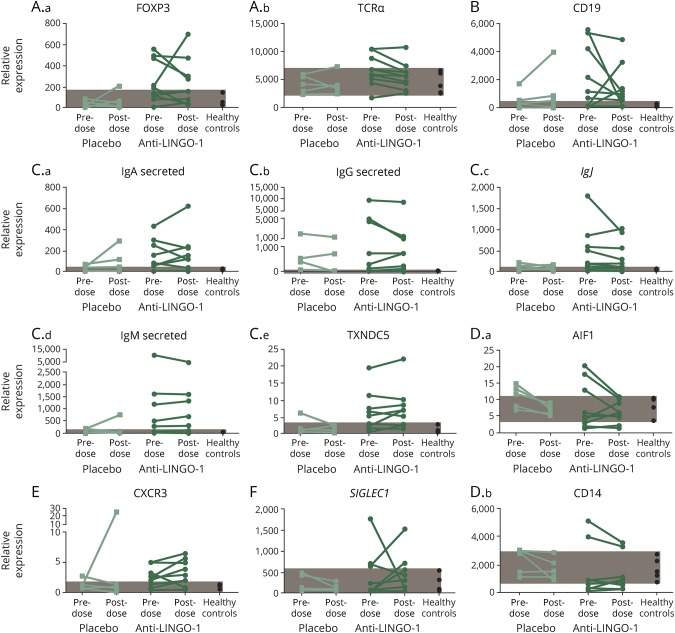
Figure 3. Gene expression in CSF cell pellets.
Gene expression in CSF cell pellets reflecting (A.a–A.b) T cells, (B) B cells, (C.a–C.e) plasma cells, (D.a–D.b) myeloid cells, (E) T-cell activation, and (F) type I IFN response before and after opicinumab or placebo treatment. Each line connects the 2 time points of an individual patient. Patients from the 30-, 60-, and 100-mg/kg groups have been combined. Healthy range (shaded area) was determined by the minimum and maximum values of 5 healthy control patients evaluated. Ig = immunoglobulin; TCRα = T-cell receptor alpha.

