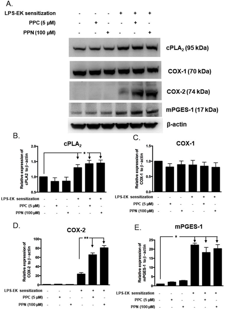
Figure 6. Western (Immuno) blot analyses for cPLA2, COX-1, COX-2 and mPGES-1 with relative expression ratios to β-actin following prior priming with LPS-EK.

pM derived from TLR4-WT mice were sensitized for 4 h with 100 ng/ml LPS-EK. Initial incubation media containing LPS-EK was removed. Cells were thoroughly rinsed once with fresh media and then incubated for 16 h with either fresh medium alone or media containing either PPC or PPN. Total cellular lysates were prepared and subjected to Western blot analysis. (A) Representative immunoblot results are shown. The histograms (B – E) represent the OD ratio of target immunoblot signal from (A) after normalization to the housekeeping protein β-actin. The experimental data are presented as the mean ± SEM from three independent experiments. * p ≤ 0.05, ** p ≤ 0.01, n = 3.
