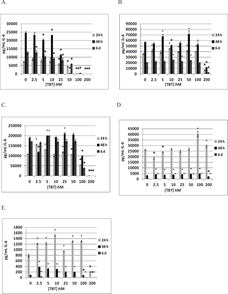
Figure 1.
Effects of 24 h, 48 h and 6 day exposures to TBT on IL-6 secretion from highly purified human NK cells, monocyte-depleted PBMCs, PBMCs, PBMCs plus granulocytes, and granulocytes in individual donors. A) NK cells exposed to 0–200 nM TBT (donor KB169). B) Monocyte-depleted PBMCs exposed to 0–200 nM TBT (donor F156). C) PBMCs exposed to 0–200 nM TBT (donor F261). D) PBMCs plus granulocytes exposed to 0–200 nM TBT (donor F261). E) Granulocytes exposed to 0–200 nM TBT (donor F261). * Indicates a significant decrease in secretion and + indicates a significant increase in secretion compared to control cells (cells treated with vehicle alone).

