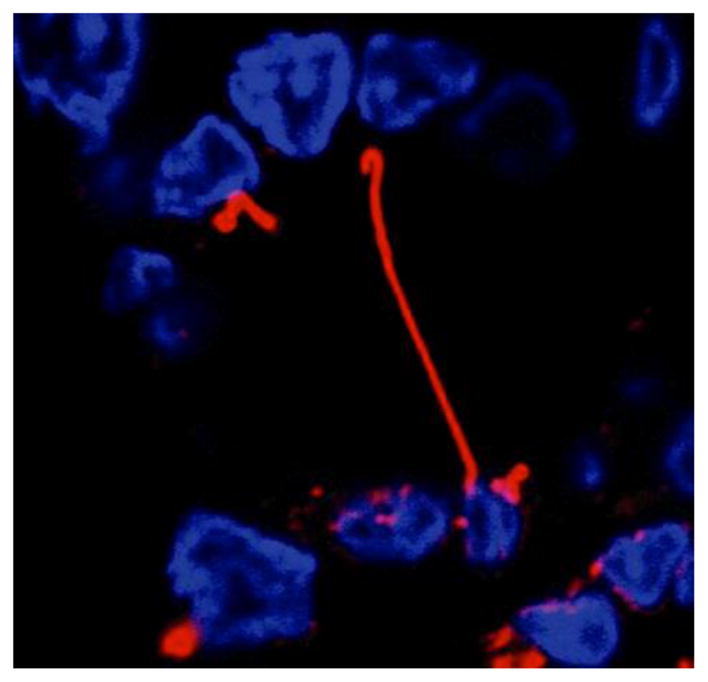
Figure 1. Primary cilium.

Confocal immunofluorescence showing a cross-section of a bile duct, where the nuclei of cholangiocytes lining the duct are stained in blue, and primary cilium extending into the ductal lumen in red.

Confocal immunofluorescence showing a cross-section of a bile duct, where the nuclei of cholangiocytes lining the duct are stained in blue, and primary cilium extending into the ductal lumen in red.