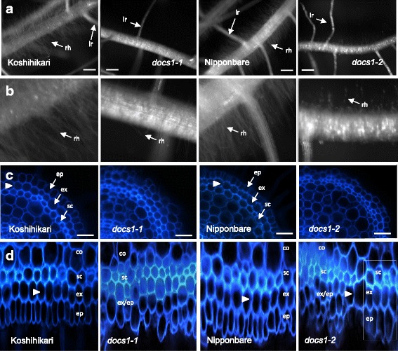
Fig. 2.

docs1–1 and docs1–2 root mutant phenotypes compared to their respective Koshihikari and Nipponbare controls. a Root hairs of docs1–1 and docs1–2 mutant roots are much shorter and less abundant than their respective controls. lr, lateral root; rh, root hairs. Bars 1 mm. b Enlarged views of the root hair zones from (a). c Organization of outer root cell layers (epidermis (ep), exodermis (ex) and sclerenchyma (sc)) are affected in docs1–1 and docs1–2 mutant roots as observed on root cross-sections. The exodermis layer is characterized by a decreased of UV fluorescence on their radial cell walls due to Casparian strip presence (white arrowheads). Bars 20 μm. d Disorganization of outer layers is highlighted on polar views of the root cross-sections. Full images of these polar views made with the imageJ software (Lartaud et al. 2015) are displayed in Additional file 1: Figure S1. The white box in the docs1–2 polar view highlights a zone where the three outer cell layers are less disorganized and where the exodermis and epidermis cell layers seem to have differentiated normally. co, cortex
