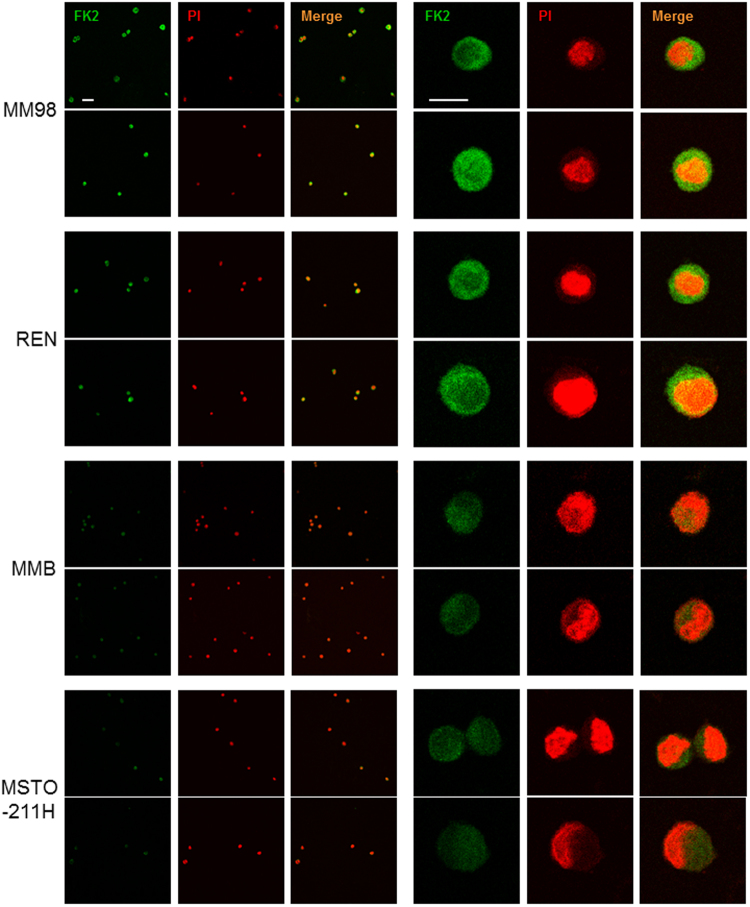
Figure 5.
Immunofluorescence microscopy of polyubiquitinated proteins in MPM cell lines. Cells were stained with FK2 antibody, which specifically recognizes poly-ubiquitin chains, and analyzed by confocal microscopy in three independent experiments. Nuclei labelled red by propidium iodide (PI); size bar: 50 μm lower magnification (left) and 10 μm higher magnification (right). Representative images. Cell area (µ2), mean ± St. dev.: MM98 108 ± 11, REN 117 ± 9, MMB 93 ± 12, MSTO-211H 110 ± 8.

