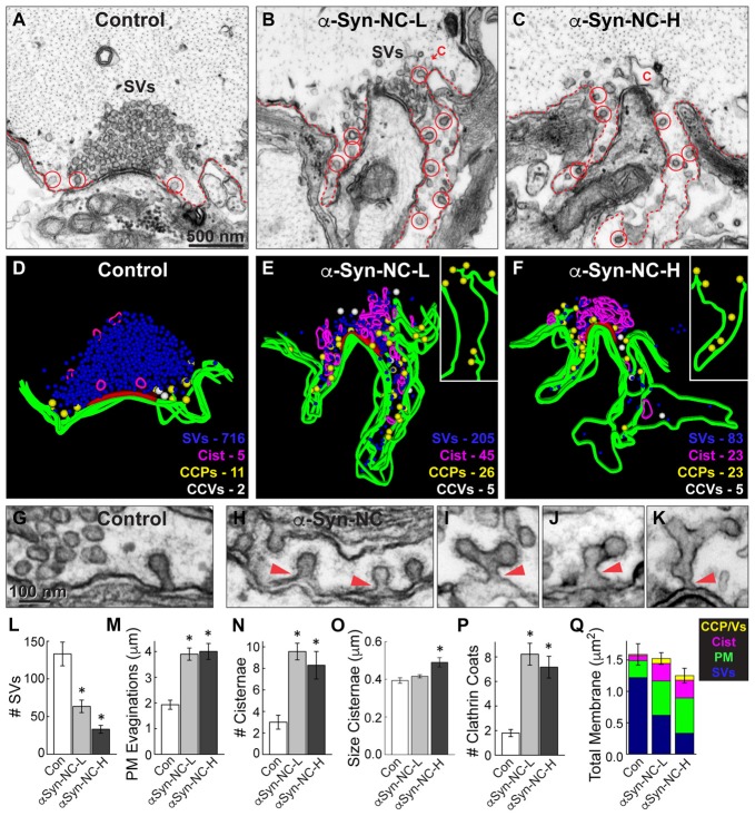
Figure 5.
α-Synuclein NC dimer also inhibits SV endocytosis. (A–C) Electron micrographs showing a control synapse and those treated with either low or high concentrations of α-synuclein NC dimer (α-Syn-NC). L, low concentration; H, high concentration. NC dimer produced a phenotype that nearly identical to that caused by CC dimer (see Figure 4). After treatment with NC dimer, synapses exhibited SV recycling defects, as shown by a reduction in SVs, expanded PM evaginations (dotted lines) and greater numbers of cisternae (C) and CCPs with constricted necks (red circles). Scale bar in (A) applies to (B,C). (D–F) 3D reconstructions show the SV recycling defects induced by NC dimer, as demonstrated by a loss of SVs (blue), larger PM evaginations (green) and build up of cisternae (magenta), CCPs (yellow) and CCVs (white). Red slab marks the active zone. (G–K) Electron micrographs show typical CCPs at control (G) and NC dimer-treated synapses (H–K). Note the abundance of CCPs, including those with complex structures induced by NC dimer. Arrowheads indicate their connections with the PM. (L–Q) Quantification of the SV recycling defects produced by NC dimer. The reduction in SVs (L) was compensated by increased PM evaginations, cisternae and CCPs and vesicles (M–Q). Data reported indicate the averages per synapse, as measured from single sections. Bars represent mean ± SEM from n = 10–24 synapses, 1–2 axons. Asterisks indicate statistical significance (p < 0.05) by ANOVA, as compared to the controls.

