Figure 2.
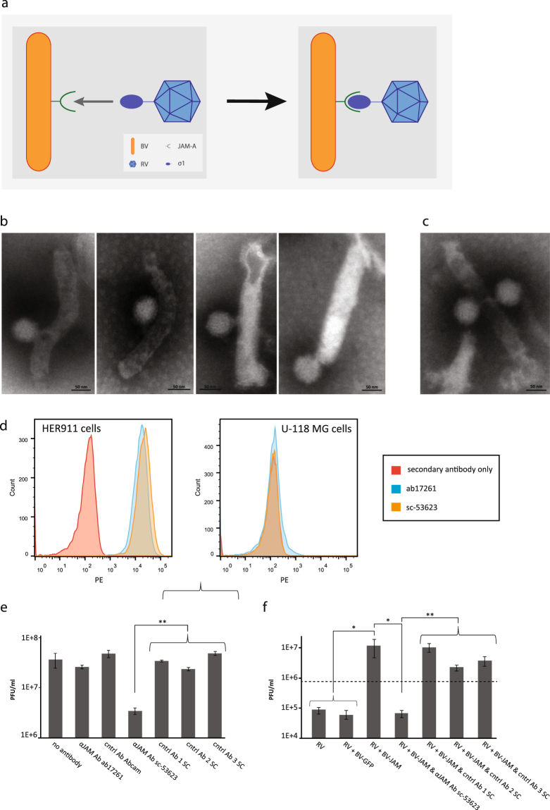
RV and BVJAM associate and form a biviral complex. (a) Schematic representation of the BVJAM-RV complex. RV attachment protein σ1 bound to JAM-A expressed on the BV envelope. (b,c) Electron microscopy images of BVJAM-RV complexes. The virions were negatively stained with uranyl acetate. Most complexes consisted of one BVJAM and one RV virion (b), some complexes showed other combinations of single or multiple BVJAM and RV virions (c). (d) Flow cytometry analyses of recognition of JAM-A on HER911 cells and U-118MG cells as negative control, by α-JAM-A antibodies ab17261 and sc-53623. (e) The RV yields from HER911 cells and culture medium upon incubation with α-JAM-A antibodies ab17261 and sc-53623 or as controls unrelated antibodies of the same provider as controls. The error bars represent the standard deviation (n = 3), p values: ** ≤ 3E-4 (f) The RV yields from U-118 MG cells and culture medium after incubation with BVJAM or BVJAM exposed to α-JAM-A antibodies sc-53623 or unrelated control antibodies of the same provider. The dashed line represents RV input. The error bars represent the standard deviation (n = 3), p values: * ≤ 4.4E-2, ** ≤ 8.8E-3.

