Fig. 3.
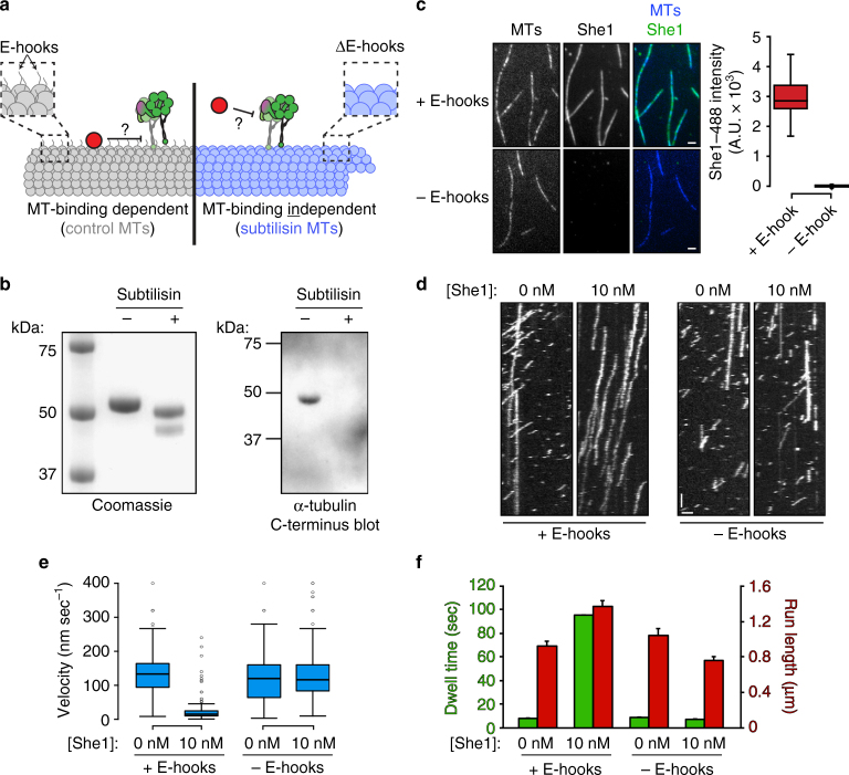
Microtubule binding by She1 is required for it to affect dynein motility. a Schematic depicting two distinct mechanisms by which She1 may affect dynein motility. She1 either requires its microtubule-binding activity to affect dynein motility (left), or it affects dynein independently of its microtubule-binding activity (right). b Coomassie-stained SDS–polyacrylamide gel (left) and immunoblot (anti-alpha-tubulin-C-terminus; right) of taxol-stabilized HiLyte647-labeled microtubules incubated with or without subtilisin, as indicated (see Methods). c 10 nM She1-TMR was incubated with either control (“+E-hooks”) or subtilisin-digested (“−E-hooks”) coverslip-immobilized HiLyte647-labeled microtubules for 5 min, then images were acquired by TIRF microscopy. Representative fluorescence images are shown (left) along with box plots of microtubule-bound She1-TMR fluorescence intensity values (scale bars, 2 µm). d Representative kymographs showing GST–dynein331 motility in the absence or presence of 10 nM She1 on either control or subtilisin-digested microtubules. Note that for each experiment in which She1 is included, She1 was pre-incubated with the microtubules for 5 min before addition of GST–dynein331, which was diluted in motility mix that also included 10 nM She1 (horizontal scale bar, 2 µm; vertical scale bar, 1 min). e Box plot of GST–dynein331 velocity values in indicated conditions. f Mean run lengths (red) and dwell times (green) for GST–dynein331 molecules along either control or subtilisin-digested microtubules in the absence or presence of She1 (error bars, standard error of the mean; n ≥ 199 individual motors for each condition). For box-whisker plots in c and e, whiskers define the range, boxes encompass 25th to 75th quartiles, lines depict the medians, and circles depict outlier values (defined as values greater than (upper quartile + 1.5 × interquartile distance), or less than (lower quartile − 1.5 × interquartile distance); Supplementary Fig. 2)

