Fig. 5.
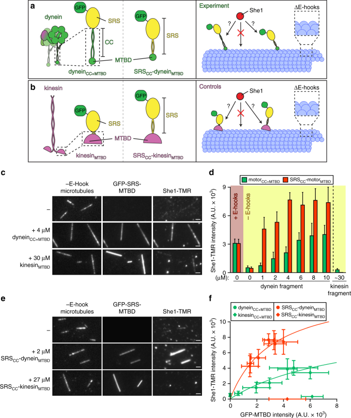
She1 binds directly to the dynein microtubule-binding domain. a–b Cartoon representations of the various GFP–seryl tRNA synthetase (SRS)–dynein (a) and kinesin (b) fusions used in the microtubule recruitment assays (left) along with a schematic of the experimental setup (right). The SRS globular domain fused to either the dynein coiled coil (CC) and microtubule-binding domain (MTBD), or the kinesin MTBD, respectively are depicted in a and b, left, while a and b, middle, depict the SRS globular and coiled coil domains fused to either the dynein or kinesin MTBD domains, respectively. c, e Representative fluorescence images of She1-TMR recruitment to subtilisin-digested microtubules by GFP–SRS–dyneinCC+MTBD (c) or GFP–SRS–SRSCC–dyneinMTBD (e), but not the respective kinesin MTBD controls. Respective images acquired from each experiment are displayed with identical brightness and contrast levels. Note that in spite of the lesser degree of microtubule-binding by the SRSCC–dyneinMTBD fusion (in e) compared to dyneinCC+MTBD (in c), more She1-TMR is recruited to microtubules by the former (scale bars, 2 µm). d Quantitation of the extent of She1-TMR recruitment to subtilisin-digested microtubules by increasing concentrations of the indicated GFP–SRS–MTBD fusion (error bars, standard deviation; n ≥ 19 microtubules, and ≥82 µm of MT length for each condition). f Relative recruitment of She1-TMR by indicated GFP–SRS–MTBD fusion. Different points reflect the mean fluorescence intensity values (along with standard deviations) for She1-TMR (fixed at 20 nM) vs. increasing concentrations of indicated GFP–SRS–MTBD fusions. Note that concentrations of the kinesinMTBD fusions were chosen such that the degree of their microtubule binding closely matched the maximal microtubule binding by the corresponding dynein fragment. As in Fig. 4e, the extent of She1-TMR microtubule recruitment was directly compared to relative microtubule binding by each GFP–SRS–MTBD fragment; Supplementary Fig. 3)

