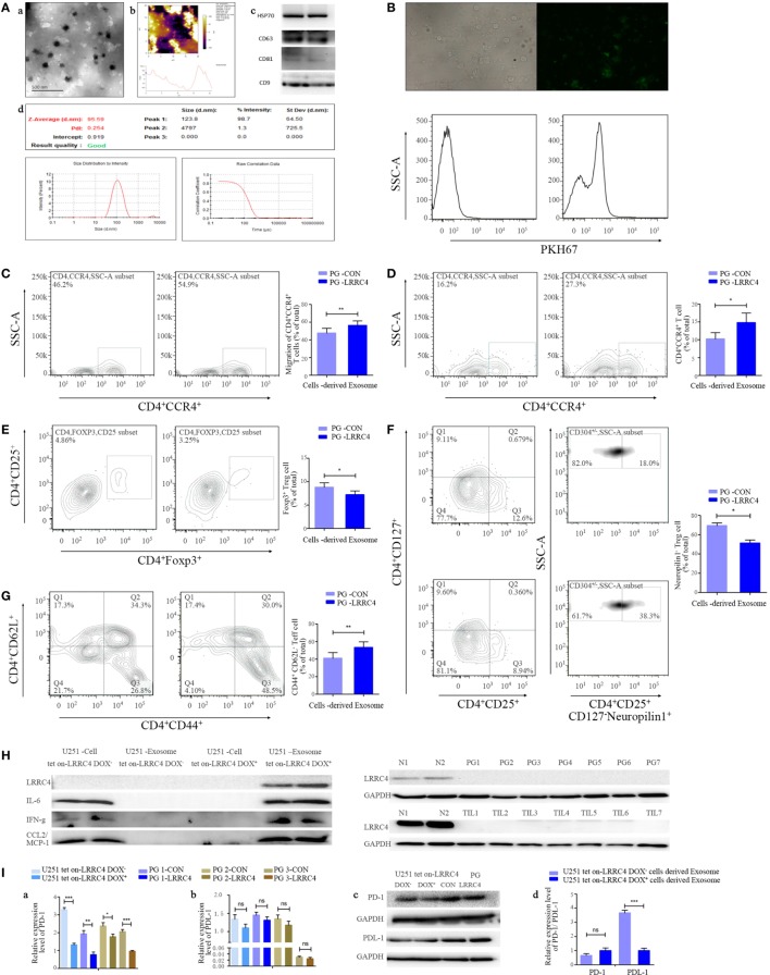
Figure 2.
LRRC4 inhibited the infiltration of Ti-Treg cells by glioblastoma multiforme (GBM) cell-derived cytokine-free and programmed cell death 1 (PD-1)-containing exosomes. [(A), a,b] Transmission electron and atomic force microscopy micrographs of exosomes (isolated from the conditioned medium of GBM cells) revealing the typical morphology and size. (c) The published exosomal markers CD63, CD81, HSP70, and CD9 were detected. (d) The particle size distribution of EVs was measured using the ZetaView® Particle Tracking Analyzer. (B) GBM cell-derived exosomes were stained with PKH67 (green) and incubated with tumor-infiltrating lymphocytes (TILs). TILs were visualized using an immunofluorescence microscope (upper panel) and FACS analysis (lower panel). (C) PG-LRRC4 and PA-LRRC4 cell-derived exosomes induced much more CD4+CCR4+ T cell chemotaxis than PG-CON and PA-CON cell-derived exosomes (**P < 0.01). (D–G) PG-LRRC4 cell-derived exosomes led to enhanced CD4+CCR4+ T cell expansion [(D), *P < 0.05], reduced CD4+CD25+Fxop3+ regulatory T (Treg) cell expansion, especially the expansion of CD4+CD25+CD127−neuropilin1− Ti-iTreg cells [(E,F) *P < 0.05], and an increased percentage of CD4+CD44+CD62L− Teff cells [(G), **P < 0.01] compared with PG-CON cell-derived exosomes; TILs isolated from GBM tissues were seeded in 48-well plates, and co-incubated with 20 μg/ml GBM cells-derived exosomes under anti-CD3/CD28 conditions. After coculturing 3 days (72 h), TILs were harvested, and CD4+CCR4+ T cells, Treg cells, and Teff cells were analyzed by FACS. (C–G). Data summarizing the results obtained for TILs generated from seven GBM patients. (H) LRRC4, interleukin-6 (IL-6), CCL2, and interferon gamma (IFN-g) protein levels were absent in U251 tet-on-LRRC4 DOX+/− cell-derived exosomes (upper panel), and no LRRC4 expression was found in PG cells from seven cases; matched TILs and normal brain tissues were from two cases (N1 and N2, lower panel). (I) PD-1 (a,c), but not PDL-1 (b,c), levels were increased in U251 Tet-on-LRRC4 DOX+ and PG-LRRC4 cells compared with U251 Tet-on-LRRC4 DOX− cells and PG-CON cells (*P < 0.05, **P < 0.01, and ***P < 0.001). (d) The expression of PDL-1, but not PD-1, was increased in U251 Tet-on-LRRC4 DOX+ cell-derived exosomes compared with U251 Tet-on-LRRC4 DOX− cell-derived exosomes (*P < 0.001).

