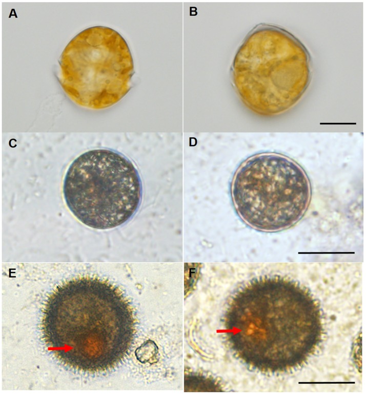
Figure 1.
Light microscopic observations for the different morphology of the vegetative cells and resting cysts of Scrippsiella trochoidea. (A,B) A typical swimming (with two flagella) vegetative cell with conical epitheca, round hypotheca, and apical process. The vegetative cells, immature cysts, and mature cysts could be clearly distinguished from morphological features under light microscope, Scale bar = 10 μm; (C,D) immature cysts (sampled from ~40-day-old culture) with a circular shape, without flagellum (thus not swimming), red body and spine. Scale bar = 20 μm; (E,F) egg or oval shaped mature resting cysts (sampled from 60-day-old culture) with a red accumulation body (arrows) and numerous surface spines. Scale bar = 20 μm.

